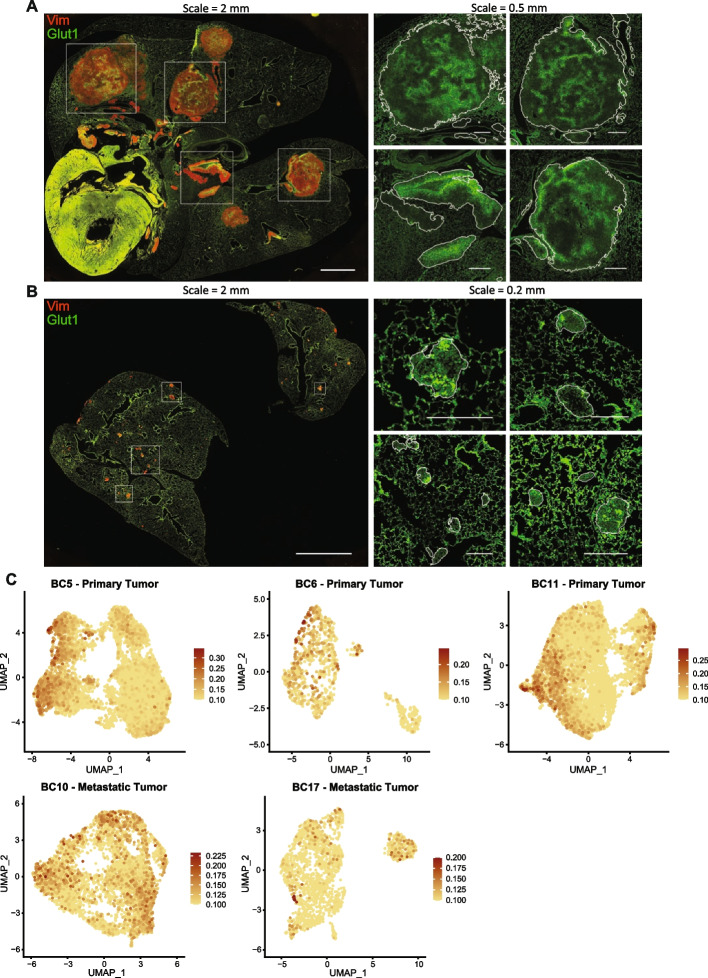
Fig. 4.
Heterogeneity in glycolysis activation identified within lung-colonizing osteosarcoma lesions. A, B Immunofluorescence staining of mouse lungs bearing OS-17 and 143B lung-colonizing tumors, respectively, for GLUT1 (green; a marker of glycolysis) and vimentin (red; marker to identify osteosarcoma cells). Magnified GLUT1 staining is shown for the boxed regions on the whole-section images. Lesion edges are indicated by white outlines in the magnified regions. Tumors showed a high degree of intra-tumor variation in GLUT1 staining intensity. C FeaturePlots for the glycolysis module score in patient primary and metastatic tumor datasets. The module score for Glycolysis was calculated using the “AddModuleScore” function in Seurat with MSigDB HALLMARK_GLYCOLYSIS genes as input features for the expression program

