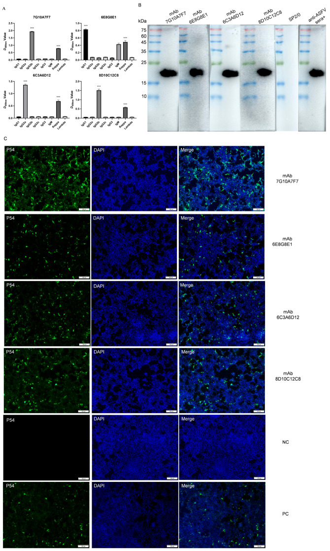
Figure 3.
Characterization of mAbs against p54: (A) isotype determination of mAbs 7G10A7F7, 6E8G8E1, 6C3A6D12, and 8D10C12C8; (B) Western blotting results of mAbs against p54 protein. SP2/0 cell supernatants and positive anti-ASFV sera (anti-ASFV sera+) were used as negative and positive controls, respectively; (C) the reactivity of mAbs was analyzed with immunofluorescence assays. Cells were fixed at 48 hpi and incubated with hybridoma supernatants as the primary antibody and FITC-conjugated goat anti-mouse IgG as the secondary antibody. Data are presented as means ± standard deviations. The differences were set to be very significant at *** p < 0.001.

