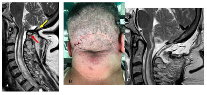
Figure 4.
This 64-year-old woman had been treated with osteodural decompression, leaving C1 intact. One year later, she was referred to us for severe nocturnal breathing disturbances and mild quadriparesis. (A) Preoperative MRI, T2-weight, sagittal view, showing a small craniectomy with C1 still maintaining a significant obliteration of the arachnoid space (red arrow). A wide syringomyelia was evident. You will notice a marked skin retraction over the craniectomy (yellow arrow). (B) Intraoperative picture showing an “arc” incision and the skin retraction. The reoperation consisted of C1 laminectomy, tonsillectomy, and dural sac augmentation. (C) Postoperative MRI, T2-weight, sagittal view showing a relatively large subarachnoid space and initial syrinx shrinkage. There was mild local cerebellar edema owing to the tonsillectomy. An asymptomatic pseudomeningocele was also evident, which resolved spontaneously within a couple of weeks. Since the first postoperative period, breathing disturbances have significantly improved, whereas quadriparesis improvement has been slower. The final CCOS score was 10.

