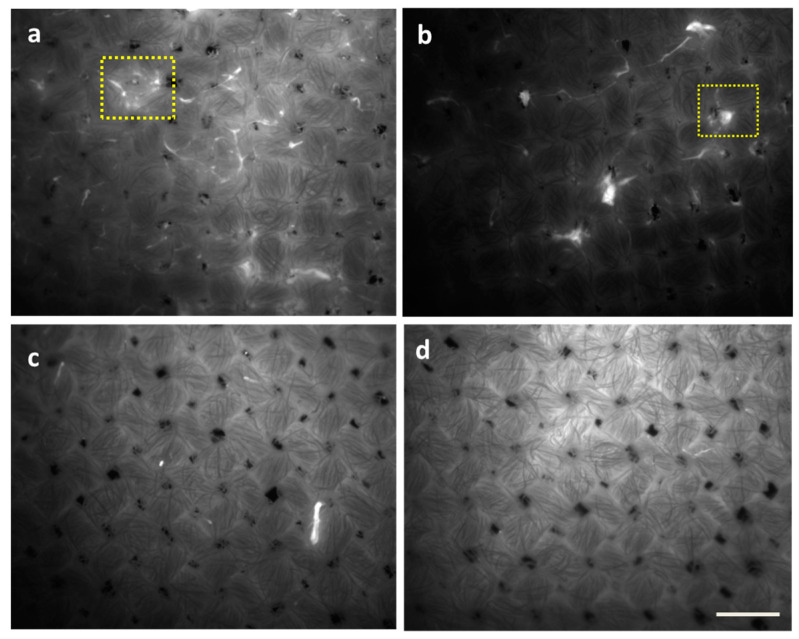
Figure 4.
Epifluorescence microscopy images of P. fluorescens grown for 48 h to form biofilms on cellulose substrates after 1 h of washing with detergent-based Formulation 1 (b), enzyme-based Formulation 2 (c), and the combination of both in F1/2 (d). Untreated biofilms are shown in (a). Bright spots (indicated by yellow boxes as examples) indicate biomass due to bacteria aggregates and biofilm attachment. Scale bar: 200 µm.

