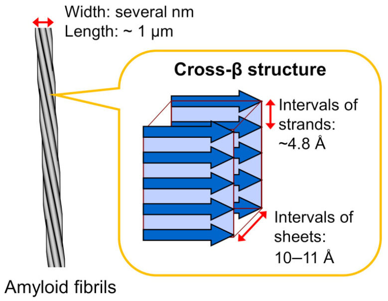
Figure 3.
Schematic illustration of an amyloid fibril. Amyloid fibrils typically show unbranched morphology, which consists of several laterally bundled protofilaments a few nanometers in width and around a micrometer in length. Protofilaments show a peculiar cross-β structure, where β-strands are stacked perpendicular to the long axis of the fibril. Reprinted with permission from Chatani E. et al., 2021 [41].

