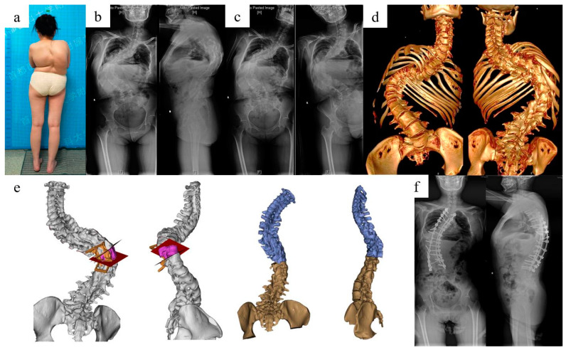
Figure 9.
Radiological images and surgical plan simulation of case 2 with kyphoscoliosis deformity. A 33-year-old female patient had a previous history of spinal correction surgery 21 years ago, and was given T10 VCR osteotomy and T3-L4 fixation and fusion utilizing the personalized surgical simulation and 3DP guidance templates. (a) Outlook for case 2; (b) preoperative full-spine standing X-ray; (c) full-spine bending X-ray; (d) full-spine CT 3D construction; (e) simulation of VCR osteotomy; (f) postoperative full-spine standing X-ray showing great improvement for the deformity.

