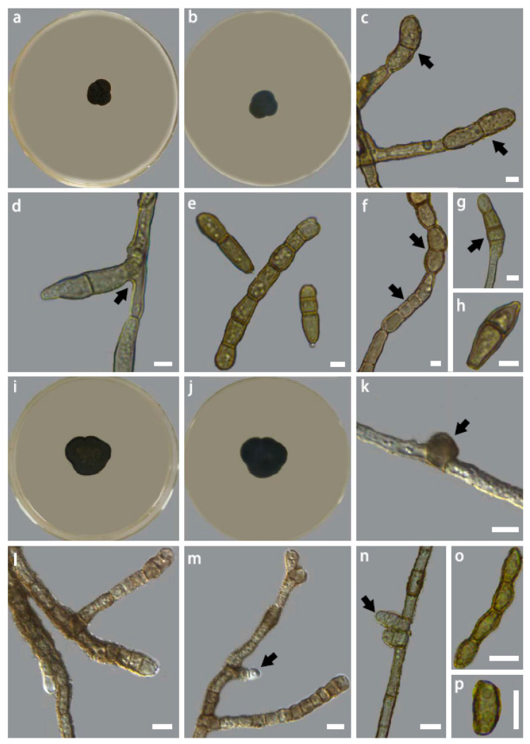
Figure 5.
Colony morphology of Intumescentia pseudolivetorum sp. nov. (CGMCC3.23635) on PDA medium after 30 days at 25 °C, (a) top, (b) reverse, (c) hyphae with thick-walled apical conidia, (d) hyphae with lateral branching with basal constriction, (e) 2, 3, and 7-celled conidia, (f) chain of conidia with both columnar and fusoid conidia, (g) apical conidia, (h) a single fusoid conidium; Colony mor-phology of Intumescentia ceratinae sp. nov. (CGMCC3.23630) on PDA medium after 30 days at 25 °C, (i) top, (j) reverse, (k) asperulous hypha with intercalary swelling, (l) hypha with variable com-partment sizes, (m,n) lateral conidial initials, (o) chain of conidia with thick walls, (p) conidium. Bars = 5μm. Morphological structures listed above are indicated with arrows.

