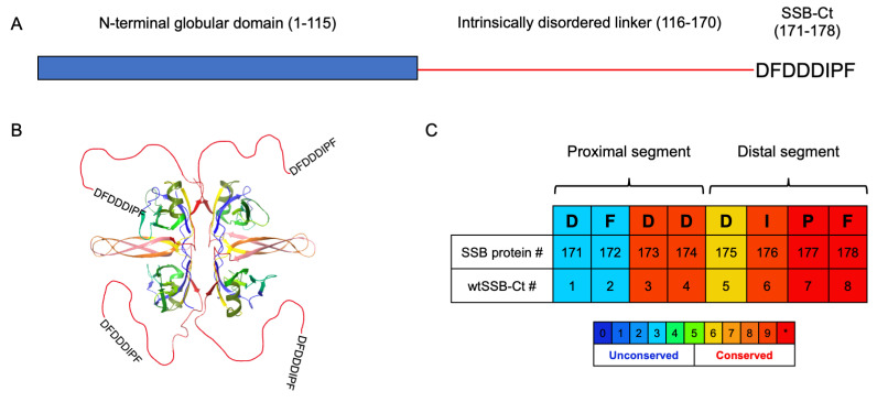
Figure 1.
(A) Domain structure of E. coli SSB protein. (B) Structure of E. coli single-stranded-DNA binding protein as a tetramer (PDB structure 4MZ9). The IDP regions are red lines, and the acidic tips are shown as amino acid sequences. (C) SSB-Ct sequence and its numbering as a segment of the whole protein. wtSSB-Ct is an octapeptide with a sequence identical to the SSB-Ct segment. Sequences are colored according to the conservation scores (see lower part of panel (C), * denotes the highest score) obtained from PRALINE [31].

