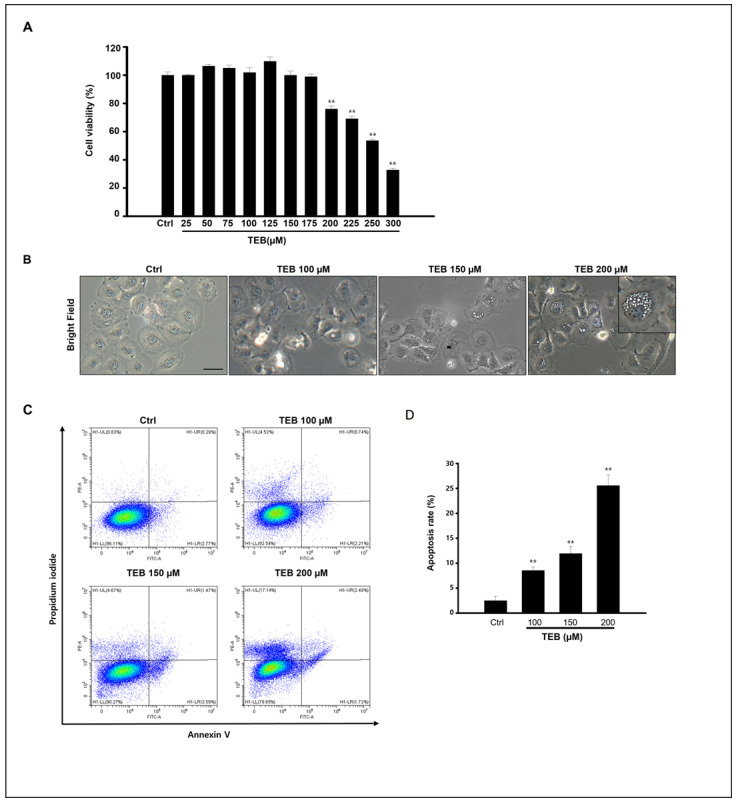
Figure 1.
Effects of TEB on viability, proliferation, and apoptosis of MAC-T cells. (A) MAC-T cell viability assessed by MTT assay. DMSO, cells treated with TEB (0–300 μM). (B) Morphological images of the cells observed under a microscope after 24 h treatment. Cells were treated with 0–200 μM of TEB in culture. Scaler bar = 20 μM. (C) The apoptotic cell death of MAC-T cells by TEB exposure was analyzed by flow cytometry at different concentrations (0–200 μM). Apoptotic cell death was determined by Annexin V-FITC/PI staining. (D) The graph shows the apoptosis rate, all data are presented as the mean ± SD of three independent experiments, and significance levels between control and treated are shown as asterisks (n = 4, ** p < 0.001).

