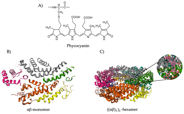
Figure 2.
Chemical, (A), and protein, (B,C) structure of phycocyanin from Spirulina platensis. (B) Represents the αβ monomer, and C) represents the three-dimensional (α3β3)2 hexamer structure of phycocyanin. The α subunit is represented by green, orange, and pink and the β subunit by grey, yellow, and brown. (C) Highlights a phycocyanobilin chromophore. α- and β- subunits have one and two phycocyanobilin, respectively. α: alpha, β: beta. Adapted from Yuan et al. (2022) [13] and Wu et al. (2016) [42].

