Figure 2.
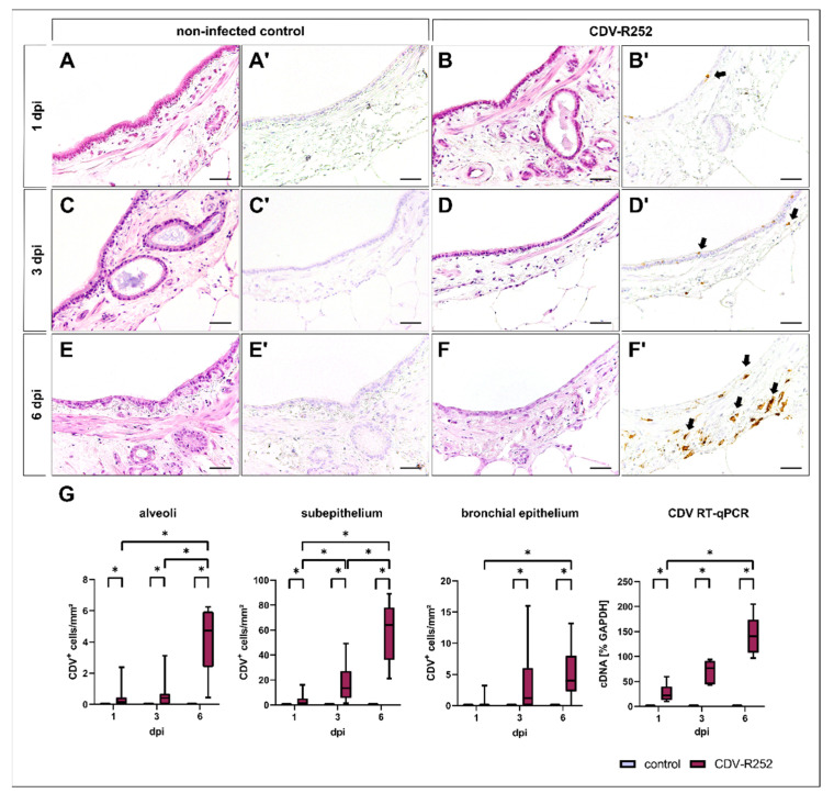
Morphology of canine precision-cut lung slices (PCLSs) and quantification of canine distemper virus (CDV) loads. Morphology remains stable during the cultivation period in noninfected (A,C,E) and CDV-infected (B,D,F) PCLSs, except from a mild attenuation of the bronchial epithelium and ciliary loss in both groups in the advanced cultivation period (E,F; HE stain). CDV antigen was not detected in control PCLSs (A’,C’,E’), while CDV-infected PCLSs showed increased viral loads during the infection period, mainly within the bronchial subepithelium (B’,D’,F’). Quantification of CDV antigen by immunohistochemistry and CDV cDNA by RT-qPCR revealed a progressive course of infection (G). Box and whisker plots show median and quartiles with maximum and minimum values. Significant changes are labelled by an asterisk (p ≤ 0.05, Kruskal–Wallis H test). dpi = days postinfection; GAPDH = glyceraldehyde 3-phosphate dehydrogenase; scale bars: 50 µm.

