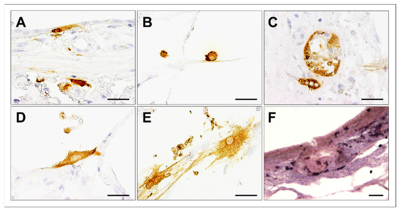
Figure 3.
Cell tropism in canine distemper virus (CDV)-infected canine precision-cut lung slices (PCLSs) at day 6 postinfection. CDV antigen was detected in bronchial epithelial cells (A), alveolar macrophages (B), bronchial glands (C), and alveolar epithelial cells (D). Occasionally, multinucleated cells containing CDV antigen were observed (E). CDV RNA was detected by in situ hybridization (dark purple, F). Scale bars: 50 µm.

