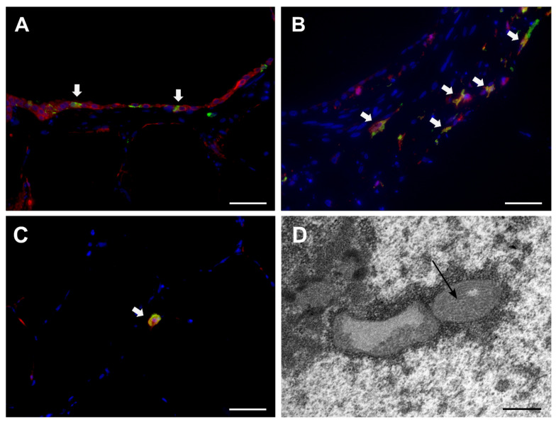
Figure 4.
Detection of canine distemper virus (CDV) in PCLSs by immunofluorescence (A–C) and transmission electron microscopy (D). Immunofluorescence double-labelling detection of CDV antigen (green), cytokeratin (red, A), or Iba-1 (red, B) revealed CDV infection of epithelial cells (white arrows, A), peribronchial histiocytes (white arrows, B), and alveolar macrophages (white arrow, C) at day 6 postinfection. Nuclear counterstaining: bizbenzimide (blue). Scale bars: 50 µm. Intranuclear inclusions of CDV nucleoprotein filaments at day 3 postinfection (D, arrow). Scale bar: 500 nm.

