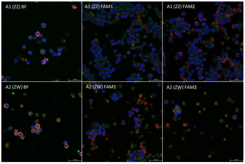
Figure 4.
The immunohistochemical staining included a stem cell-specific SSEA-1 and a germ cell-specific CVH antibody. All cells were marked with the TO-PRO-3 nucleus stain. The SSEA-1 is expressed from the cell membrane with red, the CVH from the cytoplasm with green color. The TO-PRO-3 red nucleus stain is marked blue on the confocal images.

