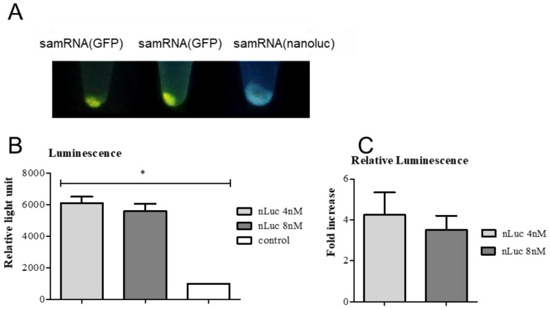
Figure 5.
Quantification of GFP and Nano-Luciferase expression demonstrated their strong production in Vero cells. In (A), pellets from Vero cells transfected with replicons expressing either GFP or nanoluc are shown under UV light (48 h after transfection). The third sample was transfected with cationic liposomes containing nLuc replicons under the same conditions. (B) Luminescence activity of Vero cells transfected with nLuc replicon complexed with 4 nM or 8 nM of cationic liposomes compared with non-transfected cells (control). The luminescence from each sample was normalized regarding the volume of the sample and protein concentration from each one. p-values are given for one-way ANOVA where * represents p < 0.005. (C) Relative luminescence of Vero cells transfected with nLuc replicon in relation to non-transfected cells. The difference in luminescence between the two samples (with a higher and lower concentration of liposomes) was not statistically significant. All experiments were performed in triplicate. Error bars show the deviations from three technical replicates.

