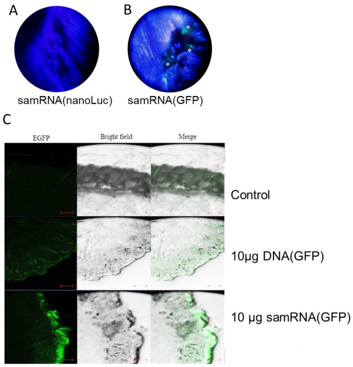
Figure 6.
Stronger GFP expression was observed after tattooing samRNA compared with plasmid DNA. Comparison of tattoo scars of animals immunized with nLuc expressing replicon (A, negative control) and GFP-expressing replicon (B), respectively, in cationic liposomes, observed under black light. Asterisks depict green spots on the skin of the animal tattooed with GFP replicons 48 h after the intradermal inoculation. Histological analyses of tissue sections (C) of BALB/c mice tattooed with liposomes encapsulating either 10 µg of DNA plasmid or RNA-replicon-encoding GFP 48 h after inoculation in the dermis. Control mice were not tattooed, and fluorescence in the control mice is due to hair in the tissue section. Images were acquired by confocal laser scanning microscopy. Data scales of 100 µm.

