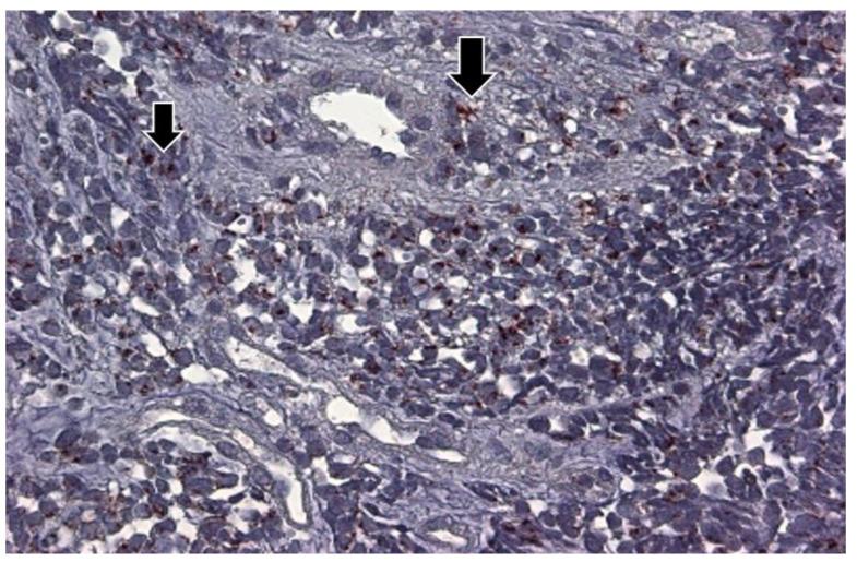
Figure 18.
The p87 immunohistochemical labeling of lung adenocarcinoma tissue at intermediate power of magnification (25×). The photomicrograph shows the adenocarcinoma diffusely involving the lung, but some normal bronchioles were observed (arrows) that exhibited a deep reddish-brown substrate, indicating the presence of p87. The dark brown p87 was observed in the cytoplasm of clearly malignant adenocarcinoma, shown on the left (smaller arrow), and in normal cells located in the upper center (large arrow).

