Figure 5.
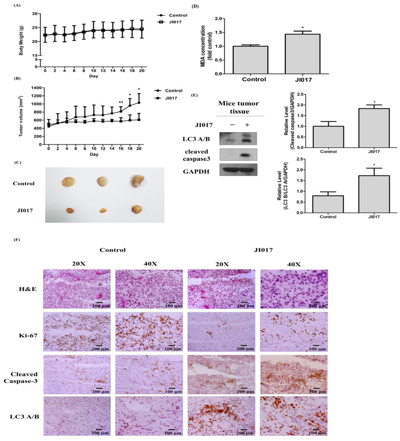
JI017 suppressed lung cancer cell growth in mice. BALB/c nude mice were subcutaneously injected with H460 cells. (A) The mouse body weight and (B) the tumor growth rate are shown. (C) Representative tumor images of the control group and the JI017-treated group. (D) The MDA concentration was assessed by a thiobarbituric reactive substance (TBARS) assay and normalized to the protein concentration. (E) Whole tissue lysates were analyzed by Western blotting with anti-LC3, anti-cleaved caspase 3, and anti-GAPDH antibodies. (F) The IHC staining of Ki-67 and cleaved caspase-3 and LC3 was carried out. Scale bar = 200 µm for 20× and Scale bar = 100 µm for 40×. The data are expressed as the mean ± SEM in all groups (n = 8–11). * p < 0.05 and ** p < 0.01 compared to the untreated group.

