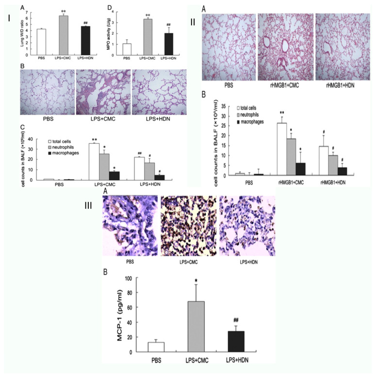
Figure 4.
Schematic representation of hesperidin reduces the acute lung damage brought on by lipopolysaccharide in mice. (I) Pre-treatment with hesperidin reduces acute lung damage brought on by LPS. (A) The lung wet to dry weight (W/D) ratio determined 24 h after LPS challenge. (B) Hematoxylin and eosin staining of lung specimens 24 h after LPS administration (H&E staining, original magnification × 100). (C) The cell counts in BALF 24 h after LPS administration. (D) The MPO activity in lung tissues 24 h after LPS challenge. ** p < 0.01 versus the PBS group, * p < 0.05 versus the PBS group; ## p < 0.01 versus the LPS + CMC group, # p < 0.05 versus the LPS + CMC group. (II) Pre-treatment with hesperidin reduces HMGB1 release and expression brought on by LPS. (A) Hematoxylin and eosin staining of lung specimens collected 24 h after rHMGB1 exposure (H&E staining, original magnification × 100). (B) The cell counts in BALF collected 4 h and 24 h after rHMGB1 exposure. The values are presented as means ± SD (n = 6–8 in each group). ** p < 0.01 versus the PBS group, * p < 0.05 versus the PBS group; # p < 0.05 versus the rHMGB1 + CMC group (III) In LPS-induced ALI, hesperidin pre-treatment reduces macrophage infiltration. (A) Lung samples were collected 24 h after LPS administration for immunohistochemistry staining of macrophage (original magnification × 400). (B) BALF samples were collected at 4 h to determine the level of MCP-1. Data are expressed as means ± SD (n = 6–8 in each group). * p < 0.05 versus the PBS group; ## p < 0.01 versus the LPS + CMC group [42].

