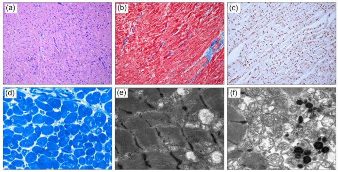
Figure 1.
Histology of the left ventricle of the explanted heart from patient A.III.2: (a) H&E-stained sections show moderate myocardial cellular hypertrophy. (b) Mild myocardial fibrosis (blue) is visualized with Masson Trichrome. (c) Immunohistochemistry with antibodies against N-Cadherin shows focal irregularity of the intercalated disks (IDs) in longitudinal sections. (d) On Richardson’s-stained resin sections, increased variation of cardiomyocytes and enlarged nuclei, partly with irregular shape, are present. (e) Representative TEM images showing disarrangement of myofibril architecture and sarcomere structure, with focal diminished and thickened Z-Bands. (f) Mitochondria are located in aggregates with swollen and dissolved cristae between myofibrils with occurrence of lipofuscin (Magnification (a–c) x100, (d) x400, (e,f) x12,000).

