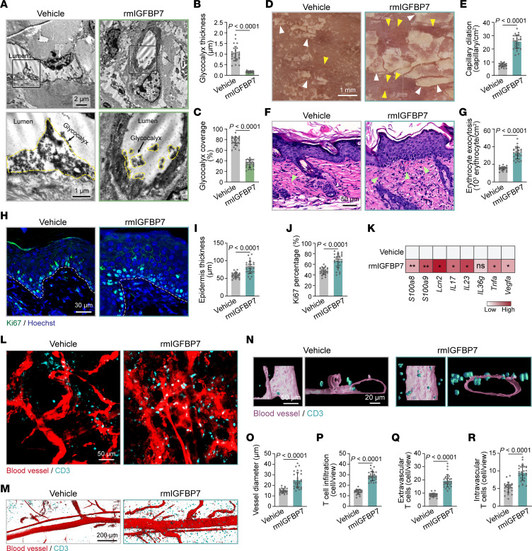
Figure 6. Elevated IGFBP7 leads to endothelial dysfunction and skin inflammation in IMQ mice.
(A) TEM of skin endothelial glycocalyx in control mice (n = 6 mice/group) that received rmIGFBP7 or vehicle i.v. The endothelial glycocalyx is highlighted by the yellow dotted lines. (B and C) Endothelial glycocalyx thickness and coverage were quantified (n = 30 vessels of 6 mice/group). (D) Dermatoscopy of IMQ mice (n = 6 mice/group) with or without rmIGFBP7 injection. Yellow arrowheads indicate dilated skin capillaries; white arrowheads indicate psoriatic scales. (E) Quantification of dilated capillaries in D (n = 30 views of 6 mice/group). (F) Skin histology of IMQ mice (n = 6 mice/group) treated with or without rmIGFBP7. Green arrowheads indicate erythrocyte exocytosis. (G) Quantification of erythrocyte exocytosis in F (n = 30 views of 6 mice/group). (H) Immunofluorescence staining for Ki67 (green) in dorsal skin from IMQ mice (n = 6 mice/group) with or without rmIGFBP7 injection. Dashed white lines mark the interface between the epidermis and dermis. (I and J) Epidermis thickness and the percentage of Ki67+ cells in the basal layer in H were quantified (n = 30 views of 6 mice/group). (K) Inflammatory gene expression in skin tissues from IMQ mice treated with or without rmIGFBP7 (n = 6 mice/group). (L) Whole-mount immunofluorescence staining for skin blood vessels (red) and CD3 (cyan) in IMQ mice (n = 6 mice/group) treated with or without rmIGFBP7. (M and N) 3D surface rendering of blood vessels and CD3 based on whole-mount immunofluorescence staining. (O–R) Quantifications of blood vessel diameter, infiltrated T cells, extravascular T cells, and intravascular T cells (n = 30 views of 6 mice/group). Data are represented as mean ± SD. Significance was calculated using unpaired Student’s t test. *P < 0.05; **P < 0.01.

