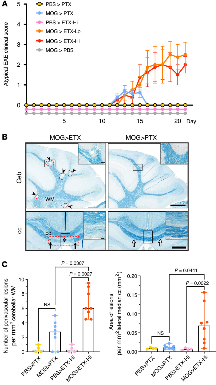
Figure 5. ETX-EAE is characterized by multifocal demyelination in the CNS.
(A) ETX-EAE mice developed atypical EAE, which is characterized by ataxia, along with classical EAE symptoms defined by ascending paralysis. Same treatment conditions as in Figure 4, CFA/MOG35-55 followed by ETX (ETX-EAE) or PTX (PTX-EAE). (B) ETX targets broader brain regions compared with PTX including cerebellum (Ceb) and corpus callosum (cc). ETX-EAE mice (left column) exhibit more focal demyelinating lesions (arrowheads and red dashed circles) in the cerebellum and corpus callosum within WM tracts when compared with PTX-EAE mice (right column). Asterisk in lower row indicates demyelinated cc. Black arrows indicate borders of a cc lesion, which is also framed with a red dashed rectangle. A corresponding location in PTX- EAE mice (right column) is indicated by white arrows. Regions framed with black boxes are shown at high magnification in inserts. (C) Quantification of lesions in the cerebellum and corpus collosum. Data in A are mean ± SEM, and data in C represent median ± range; Kruscal-Wallis test (nonparametric). n = 4 mice for PBS > PTX and PBS > ETX-Hi controls, n = 6 mice for MOG > ETX-Lo, and n = 8 mice for groups including MOG > PTX and MOG > ETX-Hi. NS, not significant. Scale bars: 1 mm in B and 100 μm in the inserts. Similar results were achieved in 2 independently repeated experiments.

