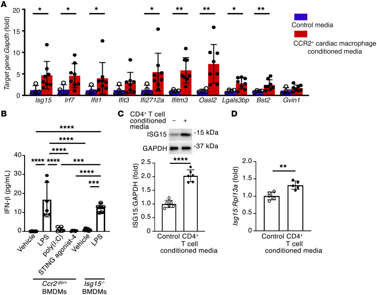
Figure 2. CCR2+ cardiac macrophages induce a cardiomyocyte IFN response.
(A) qRT-PCR for IFN response genes (Isg15, Irf7, Ifit1, Ifit3, Ifi2712a, Ifitm3, Oasl2, Lgals3bp, Bst2, Gvin1) in mouse cardiomyocytes in medium conditioned by CCR2+ cardiac macrophages isolated from Ccr2gfp/+ mouse hearts 1 week after TAC, or under control conditions. Control, n = 4; CCR2+ cardiac macrophage–conditioned medium, n = 8. (B) Culture medium IFN-β concentration in bone marrow–derived macrophages (BMDMs) from Ccr2gfp/+ mice or Isg15–/– mice incubated with LPS (1 μg/mL), poly(I:C) (500 ng/mL), or STING agonist-4 (5 μmol/L) for 24 hours (n = 6 per condition). (C and D) Immunoblotting (C; n = 6 per condition) and qRT-PCR (D; n = 5 per condition) for ISG15 in mouse cardiomyocytes exposed to CD4+ T cell–conditioned medium for 24 hours. Values are mean ± SD. *P < 0.05, **P < 0.01, ***P < 0.001, ****P < 0.0001 by unpaired 2-tailed Mann-Whitney test (A), 1-way ANOVA followed by Tukey’s post hoc test (B), or unpaired 2-tailed Student’s t test (C and D).

