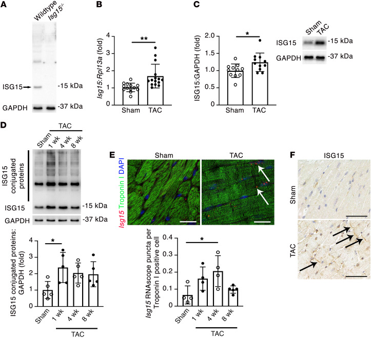
Figure 3. Cardiomyocyte ISG15 is upregulated in mouse hearts after TAC.
(A) Immunoblotting of WT and Isg15–/– mouse hearts confirming specificity of the ISG15 antibody (clone 1H9L21). (B) qRT-PCR for Isg15 in mouse hearts 8 weeks after sham or TAC. Sham, n = 13; TAC, n = 15. (C) Immunoblotting for ISG15 in mouse hearts 8 weeks after sham or TAC. Sham, n = 11; TAC, n = 11. (D) Immunoblotting for ISG15-conjugated proteins in mouse hearts 8 weeks after sham surgery or 1, 4, or 8 weeks after TAC (n = 5 per group). (E) RNAscope in situ hybridization for Isg15 and immunofluorescence for troponin I in heart sections from mice 4 weeks after sham or TAC. The arrows mark Isg15 RNAscope puncta in troponin I+ cardiomyocytes. Scale bars: 20 μm. n = 4 per group, except 8 weeks after TAC (n = 5). (F) Immunohistochemistry for ISG15 in mouse hearts 4 weeks after sham or TAC. The arrows mark positive immunostaining at, or close to, intercalated discs. Scale bars: 50 μm. Values are mean ± SD. *P < 0.05, **P < 0.01 by unpaired 2-tailed Student’s t test (B and C), or 1-way ANOVA followed by Dunnett’s post hoc test (D and E).

