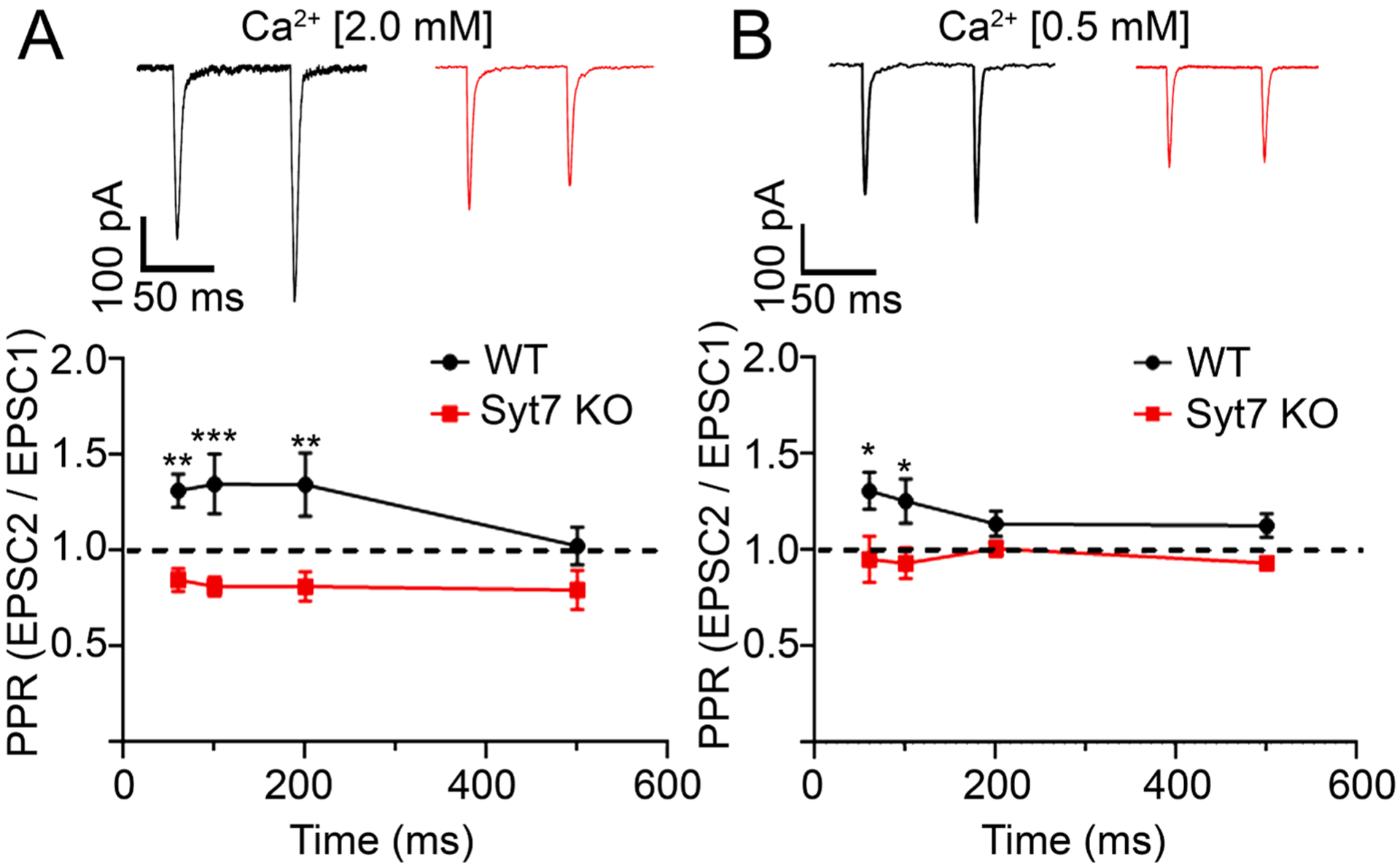
Fig. 2. Synaptic facilitation is evident in WT but not Syt7 KO medullae.

A. Representative traces (top) and averaged paired-pulse ratio (PPRs) ± SEM from evoked EPSCs at different interstimulus intervals (ISIs). PPRs at WT (black) and Syt7 KO (red) synapses are significantly different at ISIs of 60, 100 and 200 ms intervals but not at 500 ms. **p = 0.002, ***p < 0.001, Two-way ANOVA (n = 13 wt and n = 13 KO slices; >6 independent preps). B. Experiments were repeated at low extracellular Ca2+ (0.5 mM) and PPRs calculated as in A. *p = 0.01, Two-way ANOVA (n = 13 wt and n = 15 KO slices; >6 independent preps). Experiments at different calcium concentrations were performed on independent samples.
