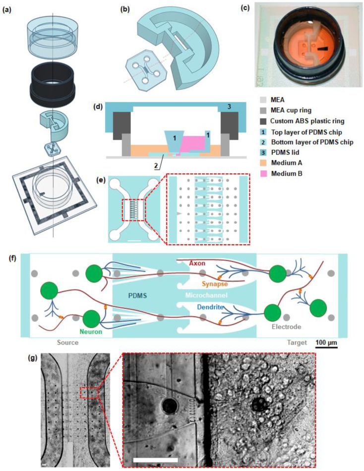
Figure 1.
PDMS chip for isolated cultures of neural cells. (a) Components and structure of the chip. From bottom to top: microelectrode array, bottom and top layer of the chip, plastic ring, PDMS lid. (b) 3D model of the bottom and top layer of the PDMS chip. (c) Photo of MEA with the chip without PDMS lid. (d) Schematic view of the MEA with the chip (side view along the line from (b)). (e) Schematic view of the microfluidic chip (scale bar 1 mm) and schematic view of the 8 microchannels of a microfluidic chip connecting two chambers. (f) Scheme of unidirectional synaptic connectivity between neural networks, formed by microfluidic chip. (g) Light field images of neural cells grown in a microfluidic chip (DIV 15). Scale bar: 100 µm.

