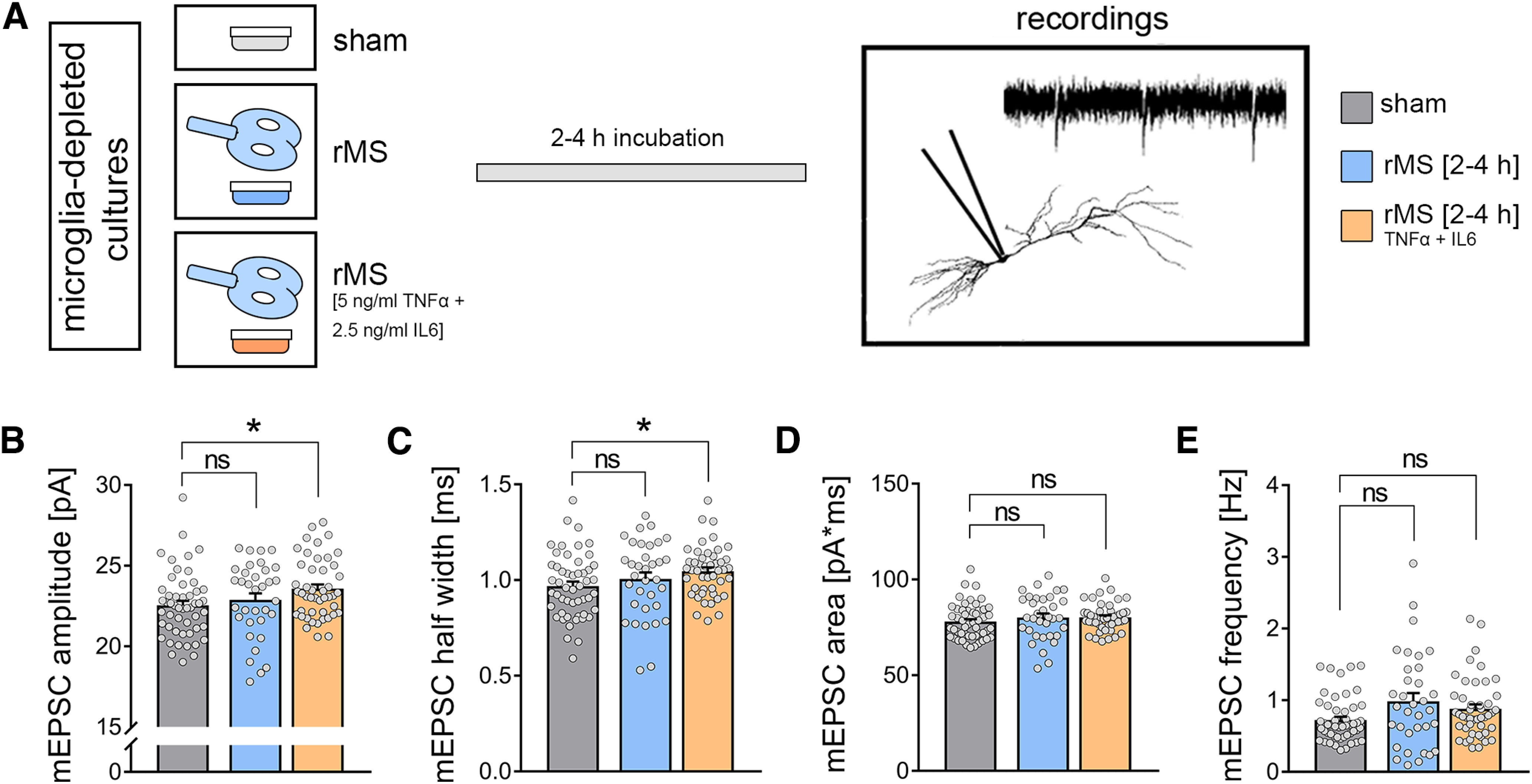
Figure 12.

Substitution of pro-inflammatory cytokines during stimulation in microglia-depleted tissue cultures rescues rMS-induced synaptic plasticity. A, Schematic of the experimental design. A subset of tissue cultures was stimulated in the presence of TNFα (5 ng/ml) and IL6 (2.5 ng/ml). Effects of TNFα and IL6 in sham-stimulated cultures are reported in the main text. B-E, Group data of AMPAR-mediated mEPSCs recorded from CA1 pyramidal neurons in sham-stimulated and 10 Hz rMS-stimulated cultures (nsham = 51 cells, nrMS = 34 cells, nrMS-TNF+IL6 = 46 cells; Kruskal–Wallis test followed by Dunn's post hoc correction). Gray dots represent individual data points. Data are mean ± SEM. *p < 0.05.
