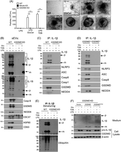
FIGURE 3.

Mincle signal-dependent release of IL-1β-containing sEVs from hepatic macrophages requires GSDMD. (A–D) immortalized murine KCs (imKCs) were transfected with small hairpin RNA to knockdown Gsdmd or Mincle and then primed overnight with 100 pg/mL LPS followed by stimulation with 20 µg/mL GluCer or 2 µg/mL TDB for 12 hours. (A) sEVs released from imKC were quantified by ZetaView and visualized by electron microscopy. (B) Western analysis of sEVs from WT and Gsdmd KD imKCs using antibodies against the indicated proteins (Rec: recombinant protein as + control; p: precursor; m: mature IL-1β or IL-18). Note: For all western using sEVs, sEVs isolated from an equal volume of cell culture media were loaded in gels; sEVs were not normalized for particle number. (C) IL-1β was immunoprecipitated from WT and Gsdmd-KD imKC culture media followed by western analyses with antibodies against the indicated proteins. (D) IL-1β was immunoprecipitated from the cell culture media from WT and Gsdmd-KD imKCs and probed with antibodies to IL-1β or ubiquitin under denaturing conditions. (E) Primary KC isolated from heterozygous littermates, and Gsdmd −/− mice were primed overnight with 100 pg/mL LPS followed by stimulation with 20 µg/mL GluCer or 2 µg/mL TDB for 12 hours. IL-1β was immunoprecipitated from culture media and analyzed by western blot as in (C). (F) Expression vectors containing WT Gsdmd or D276A mutated Gsdmd were transfected into Gsdmd-KD imKCs, then primed with 100 pg/mL LPS overnight, and stimulated with 20 µg/mL GluCer or 2 µg/mL TDB for 12 hours. Cell lysates and culture medium were collected for western analyses for indicated proteins. N=3 independent experiments. Data were represent mean±SEM. One way ANOVA ***p < 0.001. Abbreviations: GluCer, glucosylceramide; GSDMD, gasdermin D; Gsdmd-KD knocked down Gsdmd; LPS, lipopolysaccharide; Mincle-KD, knocked down Mincle; sEVs, small extracellular vesicles; TDB, trehalose-6,6-dibehenate; WT, wild-type.
