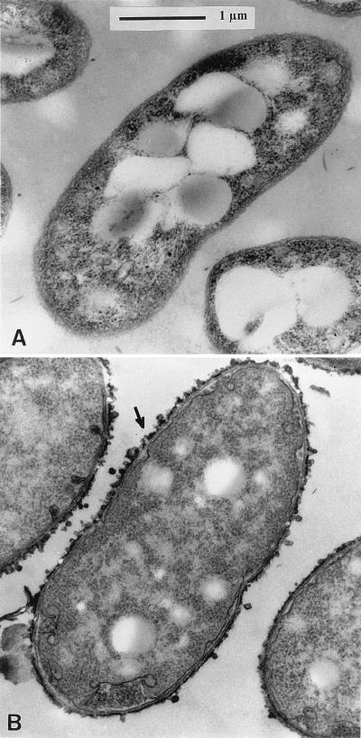
FIG. 1.
Transmission electron micrographs of transverse section of Sp7-S in free-living state, in which the outer layer of EPS is not present (A), and transverse section of Sp7-S complemented with the plasmid pAB1220-9, which shows restoration of the EPS layer (B). The arrow is pointing at the EPS layer around the cell.

