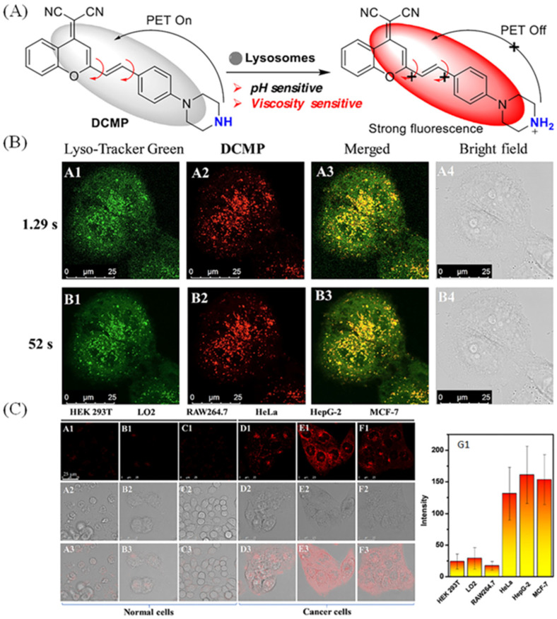
Figure 9.
(A) The design mechanisms of DCMP sensitive to viscosity. (B) Co-staining fluorescence pictures of Lyso-Tracker Green (200 nM, A1, B1) and DCMP (100 nM, A2, B2) under different scanning speed (exposure time 1.29 s and 52 s). A3, B3: the merged pictures of A1, A2 and B1, B2. (C) Fluorescent pictures of different types of cell line (cancer cells: HepG-2, HeLa, and MCF-7; normal cells: LO2, HEK 293 T, and RAW264.7) staining with DCMP (100 nM) and their relative intensity (G1) of different cells. A1–F1: confocal pictures of cells; A2–F2: DIC pictures; A3–F3: the corresponding pictures of A1–F1 and A2–F2. Reproduced from Ref. [51] with permission of the Elsevier, copyright 2022.

