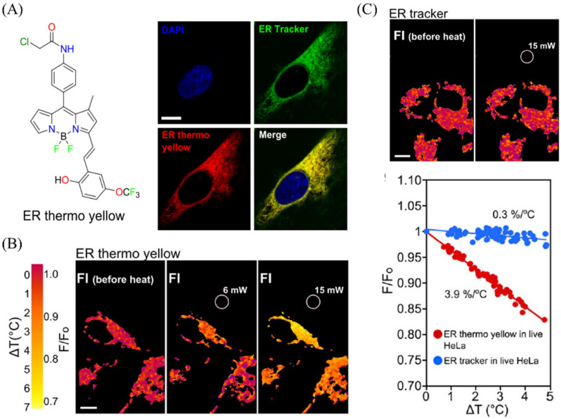
Figure 25.
(A) the molecular structure of ER probes (ER thermos yellow) for visualizing ER in HeLa cells. (B) The temperature pictures of ER thermos under diverse laser powers (6 mW,15 mW). (C) The temperature pictures of ER tracker under 15 mW laser powers, and the corresponding data. Reproduced from [120] with permission of the Springer Nature, copyright 2014.

