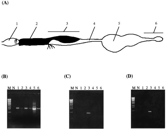
FIG. 3.
Distribution of bacteria in the gut of N. takasagoensis. (A) A schematic drawing of the gut showing the following: 1, foregut; 2, midgut; 3, mixed segment; 4, first proctodeal segment; 5, enteric valve and paunch; and 6, colon and rectum. (B) PCR using general eubacterial primers. (C) PCR using specific primers to NT-1. (D) PCR using specific primers to NT-2. M, molecular marker (λ-Eco T14I digest); N, negative control without template. The number of each lane corresponds to the numbers in panel A.

