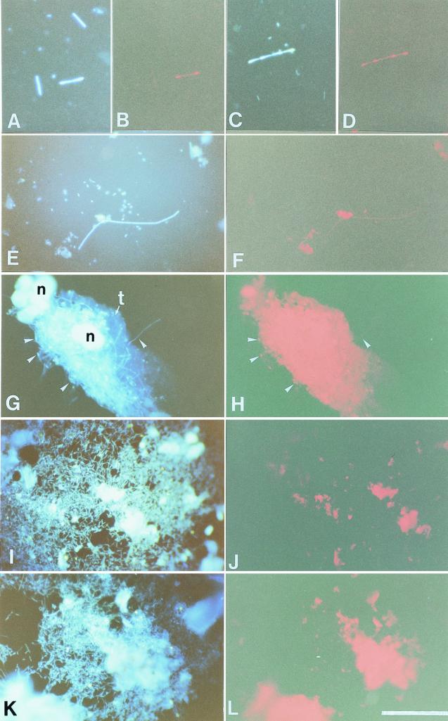
FIG. 5.
Whole-cell hybridization to 16S rRNA of the symbiotic bacteria in the mixed segment of N. takasagoensis. Rhodamine-labeled probes specific for NT-1 were applied to the gut contents, and bacteria were viewed with the DAPI filter set (A and C) and the rhodamine filter set (B and D). Stained short rods in chains have gnarl-like structure at the edges of the cells. Bacteria hybridized with the NT-2 probes were viewed with the DAPI filter set (E) or the rhodamine filter set (F). Simple short rods in chains were weakly stained, and nonspecific autofluorescence from gut contents or contaminated tissues was detected throughout the experiments. Several bacteria (arrowheads) are adhered to a piece of the mesenteric tissue (G), and some of them were detected by the NT-2 probes (H), although the strong autofluorescence from the tissue interferes with the signals from most bacteria. (I and K) Numerous bacteria were observed in the hindgut by DAPI staining. However, these bacteria are not stained with NT-1 (J) or NT-2 (L) probes. t, mesenteric tissue; n, nucleus. Bar, 50 μm.

