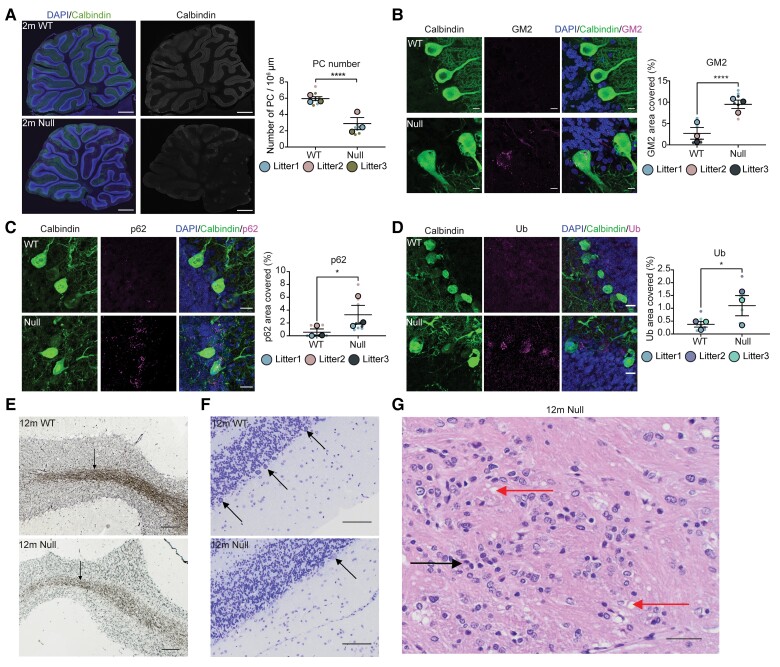
Figure 3.
Early cerebellar degeneration. (A) Sagittal cerebellar sections and quantification from 2-month-old wild-type (WT) and NHE6-null rats were stained with a Purkinje cell (PC) marker, calbindin along with DAPI. The staining of calbindin reduced in NHE6-null rats. Scale bar = 500 µm. The number of Purkinje cell cells was manually counted and divided by area (µm2). Two-tailed unpaired t-test with Welch’s correction was performed (P < 0.0001). (B) Cerebellar sections and its quantification from wild-type and NHE6-null rats at 2 months were stained with calbindin and GM2. GM2/calbindin-positive cells were detected only in NHE6-null rats. Scale bar = 10 µm. The quantification shows the per cent of area covered by GM2 (P < 0.0001). (C) Cerebellar sections were stained with p62 and calbindin. NHE6-null rats at 2 months showed increases in p62 staining (P = 0.02). (D) Cerebellar sections were stained with ubiquitin (Ub) and calbindin. The staining of Ub was increased in NHE6-null rats at 2 months (P = 0.0113). Two-tailed unpaired t-test with Welch’s correction was performed. Values from each brain section (three sections/each animal) are clustered in different colour codes according to each animal and plotted as a small dot. Means from each biological replicate are overlaid on the top of the full dataset as a bigger dot. P-value and standard error of the mean (SEM) were calculated using values from all sections from all animals (wild-type = 3, Null = 3) used in biological replicates. All data are presented as mean ± SEM. (E) Bielschowsky’s silver staining of cerebellum at 12 months. The staining in the paramedian lobule of cerebellum from NHE6-null rats prominently decreased compared to wild-type. Scale bar = 100 µm. (F) Nissl staining of paramedian lobule of cerebellum at 12 months. Arrows indicate Purkinje cell cells. The loss of Purkinje cell cells was observed in NHE6-null rats. Insets showed diffusing Nissl staining of Purkinje cell cells in NHE6-null rats. Scale bar = 100 µm. (G) Haematoxylin and eosin staining of wild-type and NHE6-null rats at 12 months. Multifocal vacuolization (red arrow) was observed in the paramedian lobule of cerebellum along with gliosis (black arrow) in NHE6-null rats. Scale bar = 100 µm.

