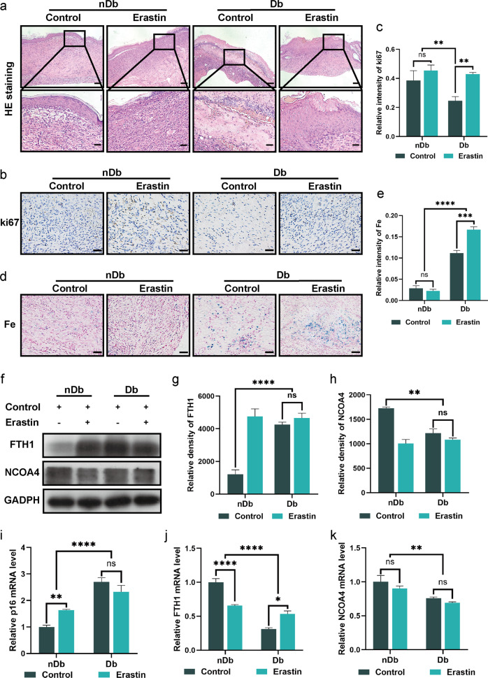
Fig. 6. Erastin accelerate wounds healing process.
a H&E staining of the wound indicated the healing situation on day 14. Scale bar = 200 μm (upper). b Immunohistochemistry staining of Ki67 in nDb and Db wound sections at day 14 after surgery. Scale bar = 50 μm. c Quantitative evaluation of the Ki67-positive cell ratio. d Prussian blue staining of the nDb and Db wounds at day 14 after surgery. Scale bar = 50 μm. e Quantitative evaluation of the Prussian blue-positive cell ratio. f Immunoblot of FTH1 and NCOA4 in wounds with the indicated treatment. g, h Quantification of FTH1 and NCOA4 immunoblotting in f. i, j The mRNA level of p16, FTH1, and NCOA4 were detected by real-time PCR in wounds with the indicated treatment. Data represent the mean ± SEM of triplicates. **P < 0.01; ***P < 0.001; ****P < 0.0001; ns nonsignificant, nDb non-diabetic, Db diabetic.

