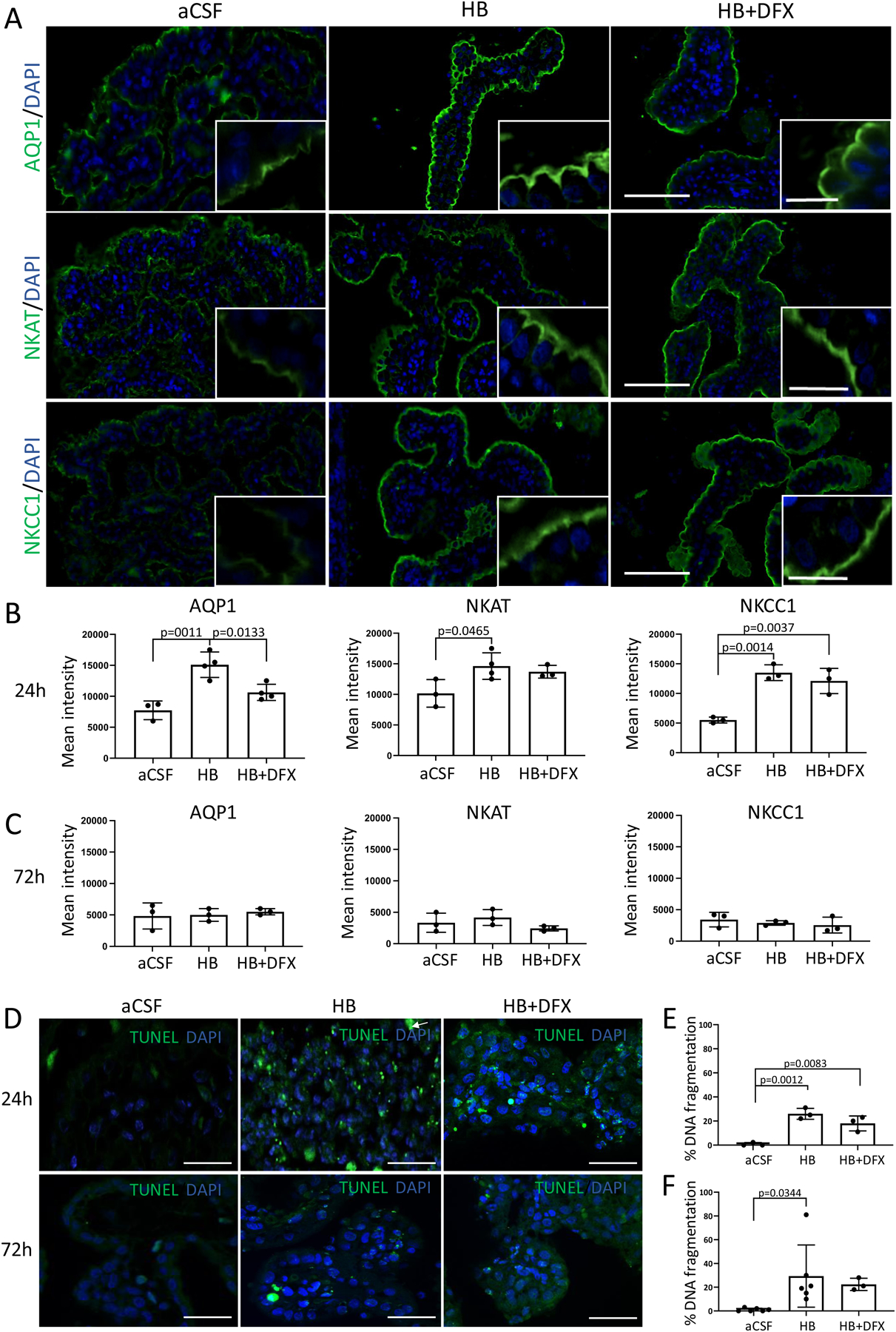
Figure 7. Hemoglobin (HB)-induced choroid plexus (ChP) expression of ion and water transporters.

(A) Representative immunofluorescence of lateral ventricle ChP aquaporin-1 (AQP1), Na+/K+/ATPase (NKAT), and Na+/K+/Cl- cotransporter 1 (NKCC1) expression 24 h after intraventricular injection of artificial CSF (aCSF), hemoglobin (HB), or HB + deferoxamine (DFX) in post-natal day 4 (P4) rats. Scale bars = 50 μm, inset scale bars = 10 μm. (B–C) Quantification of mean intensity of AQP1, NKAT, and NKCC1 expression 24 h (B) and 72 h (C) post-aCSF, HB, or HB + DFX injection showing a significant increase in AQP1 expression 24 h after IVH induction (HB injection), which was prevented by DFX treatment. Significant increases in NKAT and NKCC1 expression after IVH were not altered by DFX treatment. At 72 h post-injection, there were no significant differences in ChP expression of AQP1, NKAT, or NKCC1. Data are mean ± SEM, n = 3–4 per group; one-way ANOVA with post-hoc Tukey. (D) Representative images of right lateral ventricle ChP terminal deoxynucleotidyl transferase deoxyuridine triphosphate nick end labeling (TUNEL) assay for DNA fragmentation 24 h and 72 h after intraventricular injection. Scale bars = 50 μm. (E–F) Quantification of cellular DNA fragmentation at 24 h (E) and 72 h (F) post-aCSF, HB, or HB + DFX injection. Data are mean ± SEM, n = 3–6 per group; one-way ANOVA with post-hoc Tukey test
