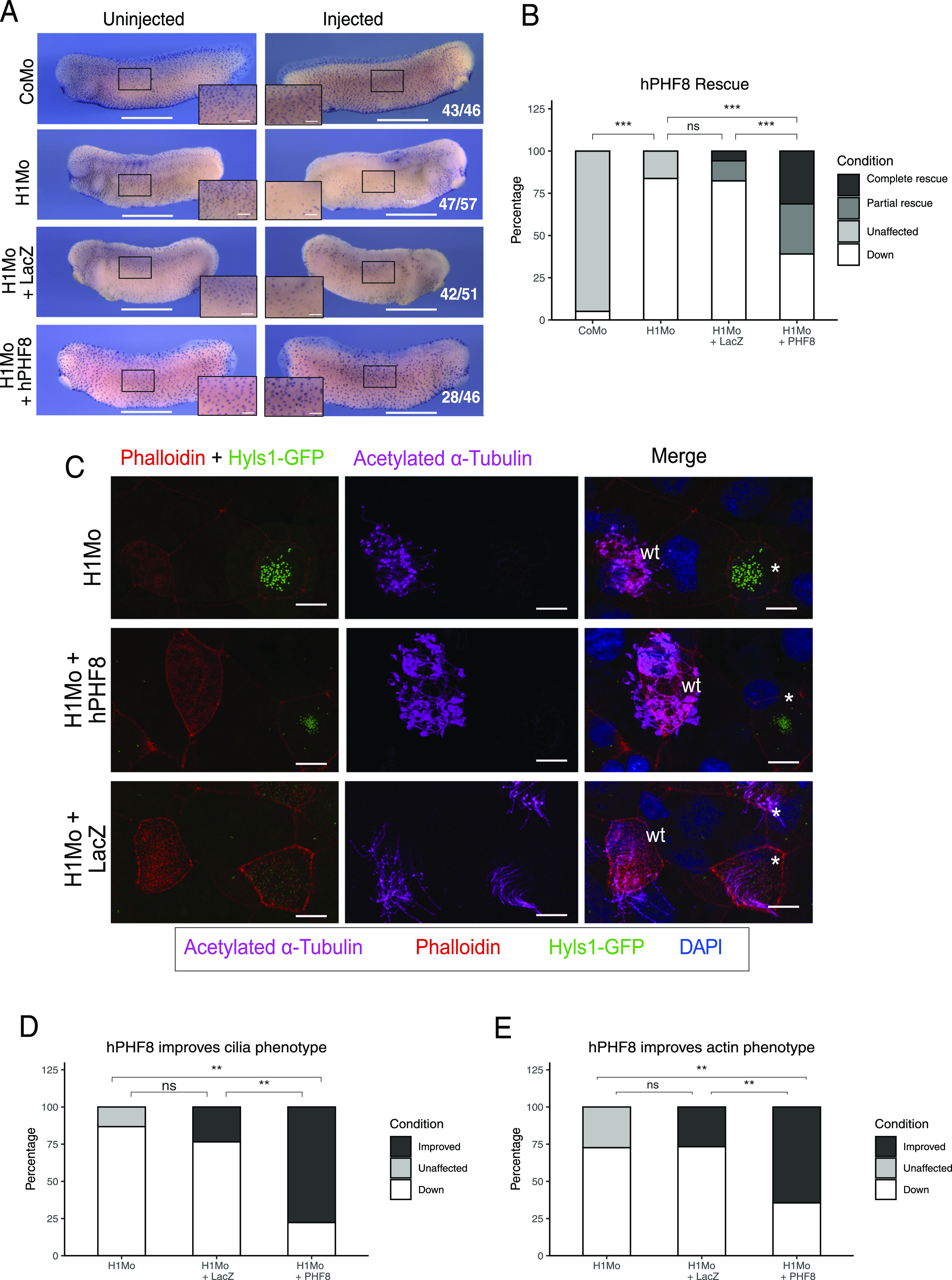
Figure 6. Rescue of ciliogenic phenotype in H1 Mo embryos with hPHF8.

(A) Representative immunocytochemistry images of the tail bud stage embryos stained for acetylated α-tubulin (multiciliated cells). Injected reagents shown on the left, uninjected, and injected sides shown on top. Scale bars = 1 mm (whole embryo) and 200 μm (inserts), n = 3 biological replicates. (B) Quantification of (A). (C) Representative confocal images detailing the multiciliated cell phenotype. Injected reagents shown on the left. The basal bodies are green (hyls1-GFP), the ciliary axonemes are magenta (acetylated α-tubulin antibody), the apical actin meshwork is red (phalloidin), and the nucleus is blue (DAPI). A mosaic injection scheme is used allowing KD and wt cells to be present in the same field of view (* = KD MCCs, wt = wildtype MCCs). Scale bar = 10 μm, n = 3 biol. replicates. (D, E) Quantification of cilia phenotype (D) and actin phenotype (E).
