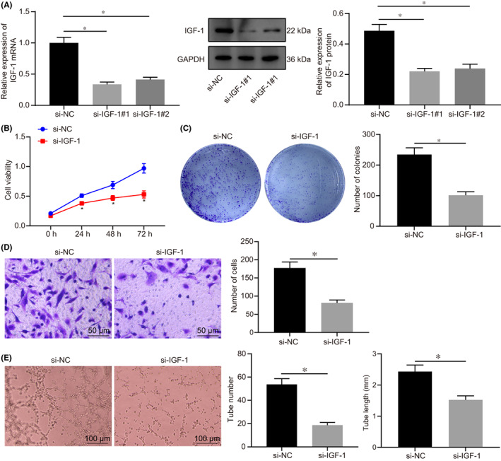
FIGURE 2.

IGF‐1 promoted proliferation and invasion of HTR‐8/SVneo cells, as well as angiogenesis in vitro. A, The expression of IGF‐1 in HTR‐8/SVneo cells after transfected with si‐IGF‐1 (1) and si‐IGF‐1 (2) determined by RT‐qPCR. B, Cell viability at 0 h, 24 h, 48 h and 72 h after transfected with si‐IGF‐1 detected by CCK‐8. C, The clonality of cells after transfected with si‐IGF‐1 detected by colony formation assay. D, The invasive ability of cells after transfected with si‐IGF‐1 detected by Transwell assay, (scale bar = 50 μm). E, Detection of lumen formation detected by HUVECs angiogenesis assay and statistical analysis of lumen number and length (× 100). All experiments were repeated three times. Data were measurement data and expressed as mean ± standard deviation. The independent sample t‐test was used for comparison between two groups. Comparisons among multiple groups were analysed by one‐way anova and followed by Tukey's post‐hoc test. * p < 0.05 vs. that of cells transfected with si‐NC. CCK‐8, cell counting kit 8; anova, analysis of variance
