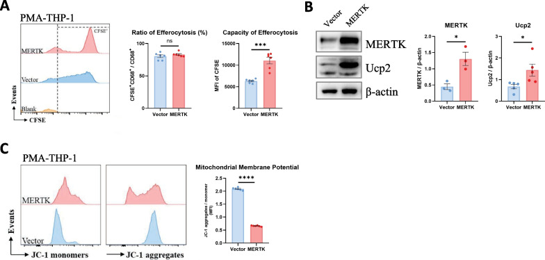
Fig. 3.
MERTK-overexpressing macrophages enhance efferocytosis function through the Ucp2/mitochondrial signalling pathway. A After coculturing apoptotic cells with vector- and MERTK-macrophages, the ratio and capacity of macrophage efferocytosis were detected by flow cytometry. Macrophages were pre-gated on CD68 + area, ratio and capacity of macrophage efferocytosis were measured by CFSE-positive ratio (CFSE + /CFSE + CD68 +) and MFI of CFSE, respectively (n = 6). B The protein expression of MERTK and Ucp2 in lung macrophages was detected by WB (n = 3–5 samples per group). C Mitochondrial membrane potential of vector- and MERTK-macrophages was measured by flow cytometry (n = 6). Data were presented as mean ± SEM. T-tests or Mann–Whitney tests were used for two-group (Vector and MERTK) comparsions. *p < 0.05; ***p < 0.001; ****p < 0.0001; n.s., not significant

