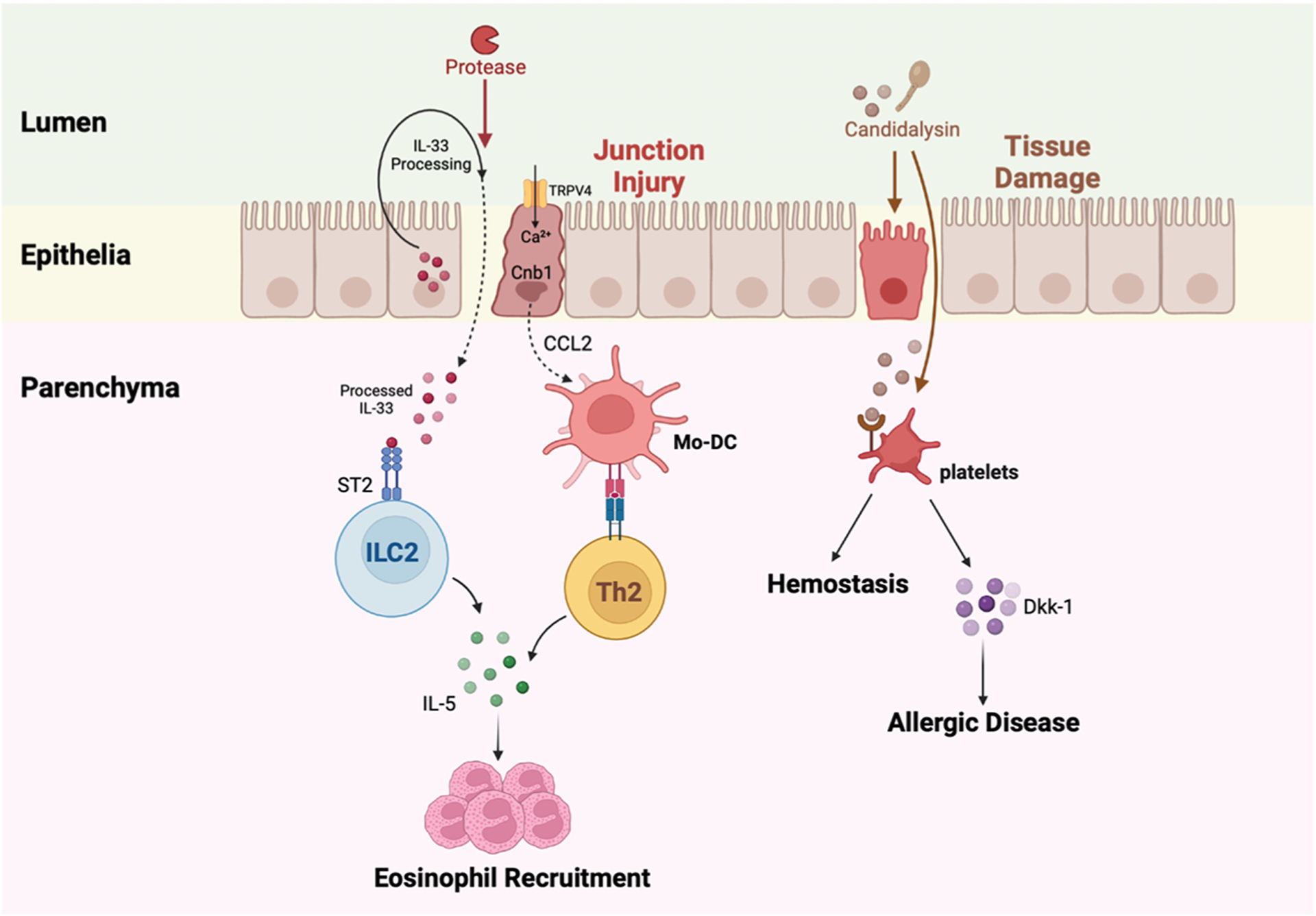
Fig. 3.

Fungal proteases and candidalysin can initiate allergenic inflammation. A fungal protease cleaves IL-33 and disrupts epithelial junctions. Processed IL-33 penetrates the barrier into the lung parenchyma, binds to ST2 receptors on type 2 innate lymphoid cells (ILC2), and drives the production of IL-5 by ILC2. This process drives eosinophil recruitment. Protease injury to the epithelial cell junctions also activates TRPV4 and causes calcium to flux into epithelial cells. Calcium signals through calcineurin and promotes CCL2 release from epithelial cells, resulting in the recruitment of Mo-DCs. Mo-DCs present antigen to T helper 2 cells in the lungs that secrete IL-5 and coordinate eosinophil influx and allergenic disease. Candidalysin, a peptide toxin released by Candida albicans, can damage lung epithelial cells in the upper airway. Platelets that arrive to limit hemorrhage interact with candidalysin, release dickkopf-1 (Dkk-1), and Dkk-1 promotes T helper 2-and T helper 17-dependent allergic responses.
