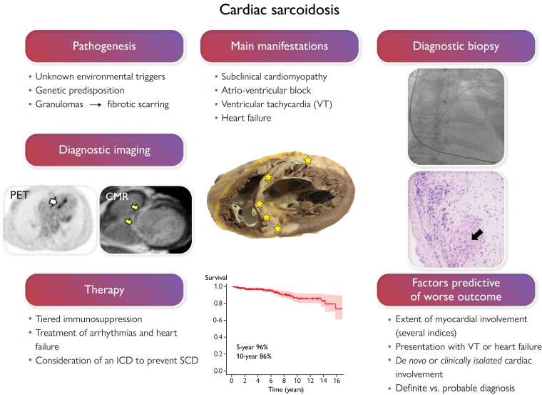
Graphical abstract.
In cardiac sarcoidosis (CS), inflammatory granulomas invade the heart leading to injury and fibrosis (yellow stars in the section of a sarcoidotic heart in the middle of the graph). CS is often subclinical, but, when clinically manifest, presents commonly with slow or fast arrhythmias or heart failure. On the left of the figure, positron emission tomography (PET) exposes focal septal uptake of 18-F fluorodeoxyglucose suggesting active inflammation (white arrow), and contrast-enhanced cardiac magnetic resonance (CMR) shows septal late gadolinium enhancement (arrows) indicating replacement fibrosis. Both constitute major diagnostic criteria for CS and entitle probable CS diagnosis if accompanied by confirmed extracardiac histology of sarcoidosis. Yet, the only way to definite diagnosis, demonstrated on the right, is myocardial biopsy showing non-necrotic granulomas (black arrow). The therapy of CS is based on immunosuppression and management of heart block, ventricular arrhythmias, and heart failure. The risk of sudden cardiac death (SCD) needs assessment and consideration of an implantable cardioverter-defibrillator (ICD). With current therapy, expected 5-year survival is well above 90% as shown by the Kaplan–Meier graph of a 398-patient Finnish CS cohort.

