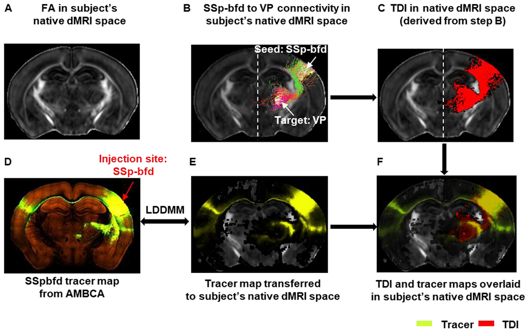
Fig. 2:

Transferring tracer map to each subject’s native dMRI space for tractography: A) Representative FA image in the native dMRI space. B) Ipsi-lateral tractogram from primary somatosensory barrel field area (SSp-bfd) to ventral posterior complex of the thalamus (VP) extracted from whole-brain tractogram shown in the native dMRI space. C) Track density image (TDI) derived from the SSp-bfd to VP streamline tractogram shown in B. D) Tract tracing image from AMBCA – selected injection site: SSp-bfd (ID:112951804). E) Tracer map transferred to the subject’s native dMRI space. F) TDI and tracer maps overlaid on subject’s native dMRI space.
