FIGURE 3.
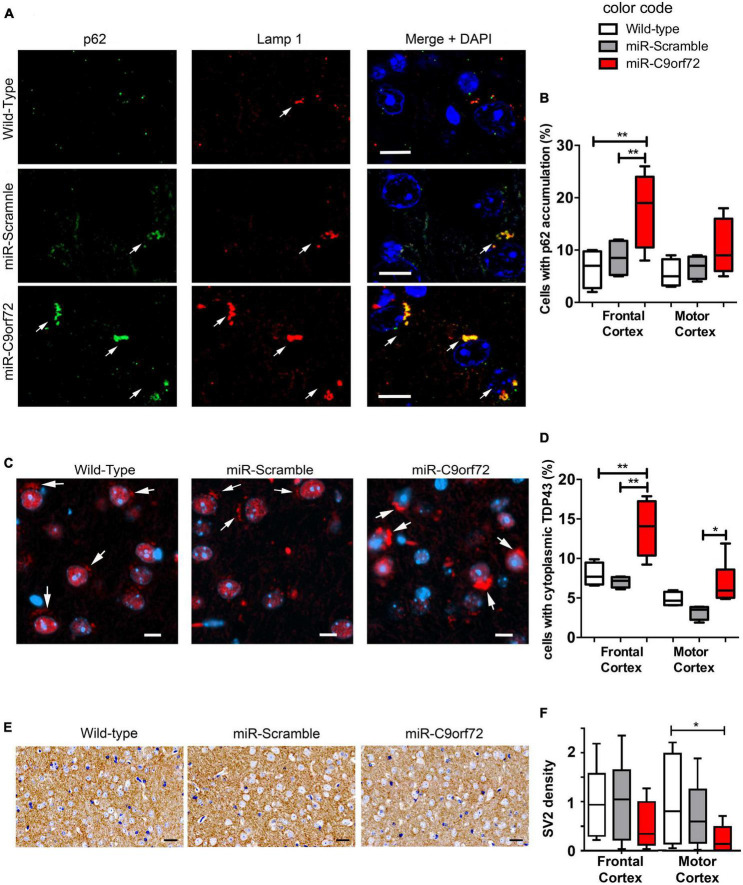
C9ORF72 knockdown causes p62 and cytoplasmic TDP-43 accumulation in the cortex. (A) Immunofluorescence co-staining of the frontal cortex from wild-type, miR-Scramble mice and miR-C9orf72 using anti-p62 (green) and anti-Lamp1 (red). Accumulation of p62 positive large structures that stain positive for Lamp1 is observed in C9ORF72 deficient mice (arrows). Scale bar: 10 μm. (B) Quantification of cells presenting p62 accumulation in the frontal and the motor cortex of controls and C9ORF72 deficient animals. (C) Immunofluorescence staining of TDP-43 (red) in sagittal sections of the cortex from wild-type, miR-Scramble mice and miR-C9orf72. Cytoplasmic structures that stain positive for TDP-43 are indicated by arrows. Scale bar: 10 μm. (D) Quantification of cells presenting cytoplasmic TDP-43 accumulation in the frontal and the motor cortex of controls and C9ORF72 deficient animals. (E) 3,3′-diaminobenzidine (DAB) staining of SV2 positive synapses in in sagittal sections of the cortex from wild-type, miR-Scramble mice, and miR-C9orf72. Scale bar: 20 μm. (F) Quantification of the normalized density of SV2 staining in the frontal and the motor cortex of controls and C9ORF72 deficient animals (the average density of SV2 staining in wild-type animals was used to nomalize all densities). For all experiments, wild-type and miR-Scramble n = 4; miR-C9orf72 n = 6. Error bars represent SEM; *p < 0. 05, **p < 0.01.

