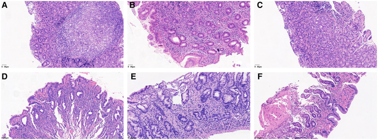
Figure 3.
He staining results showed the degree of lymphocyte cells, plasma cells and neutrophils in the gastric mucosa. (A) Lymphocytes and plasma cells accumulated in the epithelial cells and lamina propria of the gastric mucosa (severe). (B) Small amounts of lymphocytes and plasma cells were found in the gastric mucosal epithelium, and considerable amounts were found in the lamina propria (moderate). (C) Small amounts of lymphocytes and plasma cells were found in the epithelium and lamina propria of the gastric mucosa (mild). (D) Neutrophils accumulated in the epithelial cell layer and lamina propria of the gastric mucosa (severe). (E) Small amounts of neutrophils were found in the gastric mucosal epithelial cell layer, while considerable amounts were found in the lamina propria (moderate). (F) Small amounts of neutrophils were found in the epithelial cell layer and lamina propria of the gastric mucosa (mild).

