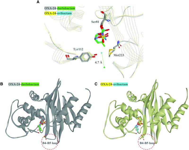
Figure 1.
Co-crystal structures of durlobactam or avibactam and OXA-24. A, The crystal structures of OXA-24 (gray) with durlobactam (green) (Protein Data Bank [PDB]: 6MPQ) and OXA-24 (yellow) with avibactam (cyan) (PDB: 4WM9) show that the water molecule in the OXA-24–durlobactam structure (green ball) is 4.7Å away from the hydrophobic bridge formed by Tyr112 and Met223 compared to the OXA-24–avibactam structure (cyanball). This difference is likely due to the methyl side chain present on durlobactam that is lacking on avibactam. B, The crystal structure of OXA-24 (gray) with durlobactam (green) (PDB: 6MPQ) reveals that the B4-B5 loop is resolved in the structure. C, The crystal structure of OXA-24 (yellow) with avibactam (cyan) (PDB: 4WM9) lacks resolution of the B4–B5 loop, suggesting that this region is flexible.

