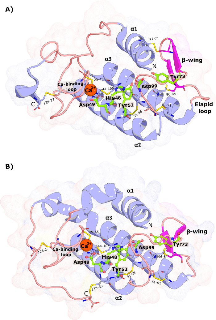
Figure 3.
| (A) Structure of the Chinese cobra svPLA2-IA (PDB 1POA) and (B) the Indian saw-scaled viper svPLA2-IIA (PDB 1OZ6). The active site residues (His48, Asp49, Tyr52, Tyr73, and Asp99) are shown as green sticks, the Ca2+ as an orange sphere, and the disulfide bonds as yellow lines. N- and C-terminal regions are also identified. The similarity in the folding is evident. The PyMOL39 molecular graphics software package was used to generate the representations.

