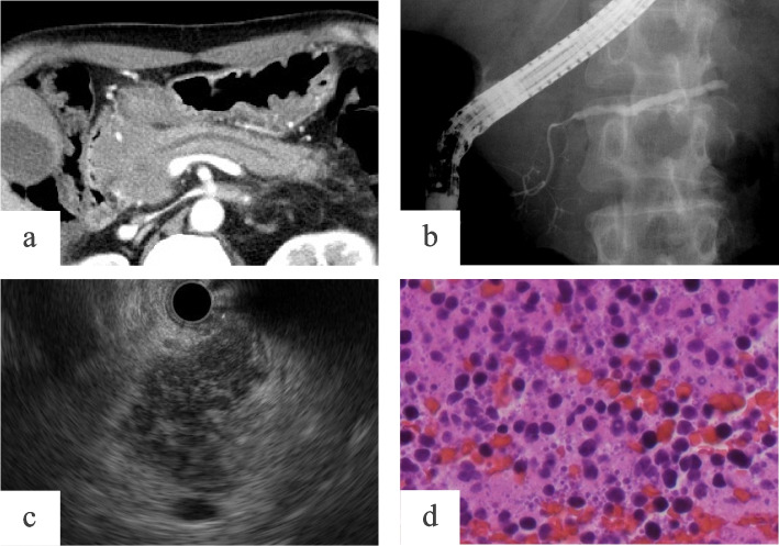
Fig. 3.
Case 2.1: A case of pancreatic lymphoma that was not definitively diagnosed from specimens obtained by endoscopic ultrasonography–guided fine-needle aspiration. A 65-year-old man was admitted to our hospital with acute pancreatitis. Contrast-enhanced computed tomography showed a homogeneous mass in the pancreatic head measuring 55 mm in maximum diameter and slight dilation of the caudal main pancreatic duct (MPD) at 3.42 mm (a). In endoscopic retrograde pancreatography, the MPD localized in the tumor was visible and narrowing smoothly, and the caudal MPD was mildly dilated (b). Endoscopic ultrasonography found a 50 mm round hypoechoic tumor with a clear boundary and scattered high echoic spots (c) suggesting autoimmune pancreatitis. Cytologic assessment of the tumor using EUS-FNA revealed atypical lymphocyte accumulation suggesting malignant lymphoma (d)

