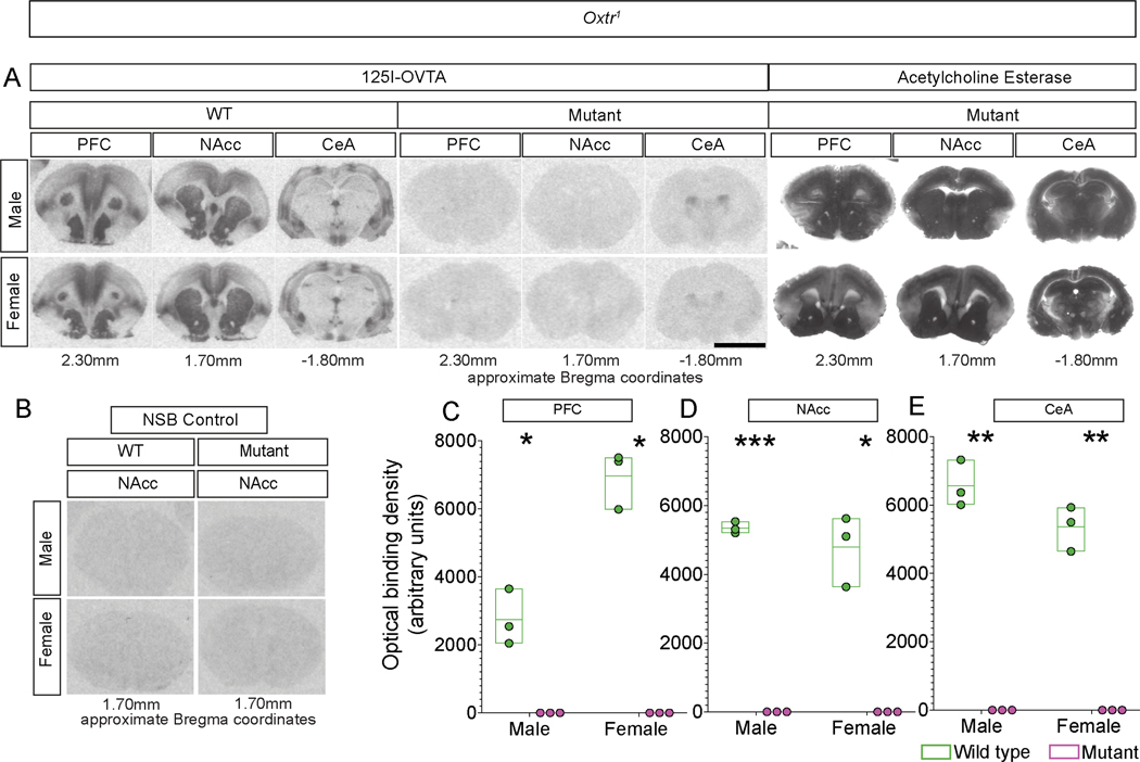
Figure 2. Oxtr mutant voles lack functional, ligand-binding Oxtr.
A. Loss of binding with the competitive agonist 125I-OVTA visualized in coronal sections through the rostral telencephalon: Left: Labeling in PFC (prefrontal cortex), NAcc (nucleus accumbens), and CeA (central amygdala) of WT sibling controls; Middle: Labeling in equivalent sections through PFC, Nacc, and CeA from Oxtr1 homozygous mutant voles; Right: The same mutant sections as in the middle panels, stained for Acetylcholine esterase to demonstrate equivalence of sections chosen for WT and mutants.
B. Non-specific binding (NSB) control shows no off-target binding in WT or Oxtr1 mutant sections.
C-E. Optical density-based quantification of binding to 125I-OVTA shows that binding is essentially undetectable in mutants null for Oxtr1 in PFC (C), NAcc (D), or CeA (E). Scale bar =5 mm; boxplot depicts max-minScale bar =5 mm; boxplot depicts max-min, midline denotes mean; n=3 for WT and mutant males and females (C-E). Scale bar =5 mm; boxplot depicts max-minSee also Figure S2.

