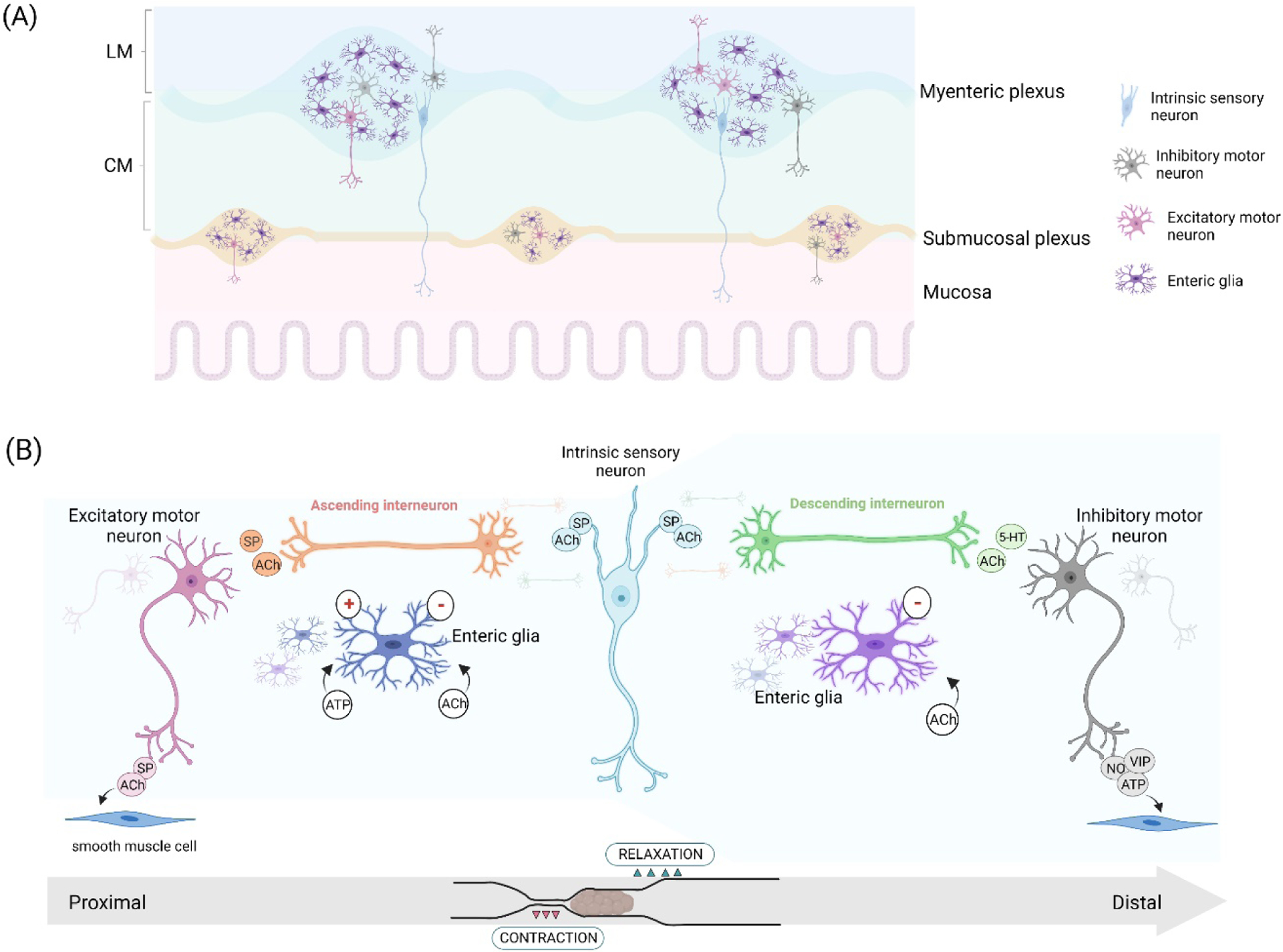
Figure 1.

Neural circuitry for controlling intestinal motor activity. (A) The basic structure of the enteric nervous system. Enteric neurons and enteric glia are found widely distributed among gut layers, notably in the nervous plexus. Inside the ganglia, multiple connections are established as well as between ganglia. (B) In the myenteric plexus, a synaptic arrangement spatially signals the contraction and relaxation of circular smooth muscle. Intrinsic sensory neurons activate ascending and descending interneurons, which in turn activate excitatory motor neurons orally and inhibitory motor neurons anally, creating coordinated motor activity for the propulsion of intestinal contents. Enteric glia signaling evoked by acetylcholine and purines reinforces pro-contractile activity and refines gut motility commands. LM=longitudinal muscle; CM=circular muscle. SP=substance P, 5-HT=serotonin, ATP=adenosine triphosphate, ACh=acetylcholine, NO=nitric oxide, VIP=vasoactive intestinal peptide.
