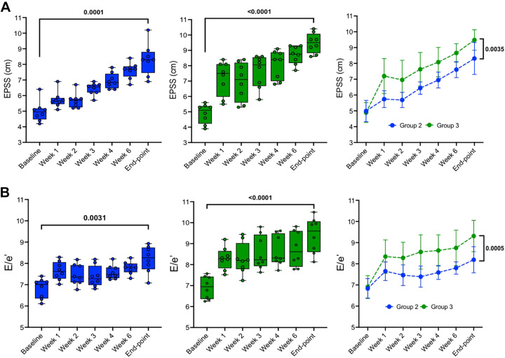
Figure 7.
Left ventricular function data during ischemic heart failure (HF) progression. A: end-point septal separation (EPSS) data acquired in M-mode from a right parasternal long-axis view. B: E/e′ data acquired and quantified through pulse wave (PW) [tissue-Doppler imagining (TDI)] images from a 4-chamber apical view. Means ± SD of each group at same time point: group 2 (n = 8; left), group 3 (n = 8; middle), and two-tailed paired t tests (right) were used to compare data between two time points (baseline vs. end point) within the same group (left and middle). Two-way ANOVA mixed model (right) was conducted to analyze time effect, groups effect and time/groups interaction effect on EPSS (P < 0.0001, P < 0.0001, and P = 0.1253, respectively), and E/e′ (P < 0.0001, P < 0.0001, and P = 0.2021, respectively). Post hoc tests with Bonferroni correction are shown at end points between the two groups.

